Difference between revisions of "SOCR Data N46 TBI ROI Volumes"
(Created page with "== SOCR Datasets - Acute and Chronic Traumatic Brain Injury (TBI) Neuroimaging study == ==Data Overview== Image:SOCR_Data_ID_April2011_NI_Pain_Fig1.gif|350...") |
|||
| Line 5: | Line 5: | ||
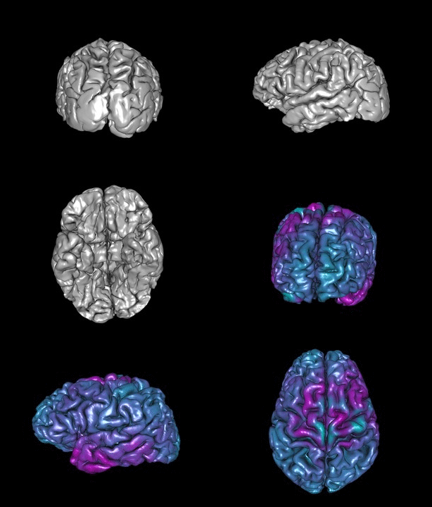
Examine the precise nature of localized brain atrophy in Traumatic Brain Injury (TBI) using non-invasive imaging methods may provide predictive biomarkers to identify patients at risk to develop Posttraumatic Epilepsy (PTE). Automated shape and volume analysis technique can disclose the presence of focal alteration in patients’ thalami after acute Traumatic Brain Injury. | Examine the precise nature of localized brain atrophy in Traumatic Brain Injury (TBI) using non-invasive imaging methods may provide predictive biomarkers to identify patients at risk to develop Posttraumatic Epilepsy (PTE). Automated shape and volume analysis technique can disclose the presence of focal alteration in patients’ thalami after acute Traumatic Brain Injury. | ||
| − | ==Data Description== | + | ===Data Description and Variable Specification=== |
[[SMHS_MissingData|missing data]] were imputed using R's MI package. | [[SMHS_MissingData|missing data]] were imputed using R's MI package. | ||
| − | + | ==Dataset== | |
| − | ==Demographic and Clinical Data== | + | ===Demographic and Clinical Data=== |
<center> | <center> | ||
{| class="wikitable" style="text-align:center;" border="2" | {| class="wikitable" style="text-align:center;" border="2" | ||
|- | |- | ||
| − | ! | + | !rowspan="2"|ID || colspan="2"|DEMOGRAPHICS ||colspan="4"|GCS||colspan="2"|GOSE||colspan="3"|INJURY_CHARACTERISTICS||colspan="3"|PERSYT_SPIKE_DETECTION||colspan="3"|SEIZURES |
| + | |- | ||
| + | |age||sex(m0f1)||field.gcs||er.gcs||icu.gcs||worst.gcs||X6m.gose||X2013.gose||skull.fx||temp.injury||surgery||spikes.hr||min.hr||max.hr||acute.sz||late.sz||ever.sz | ||
|- | |- | ||
|1||19||0||10||10||10||10||5||5||0||1||1||31||18||329||1||1||1 | |1||19||0||10||10||10||10||5||5||0||1||1||31||18||329||1||1||1 | ||
| Line 101: | Line 103: | ||
|}</center> | |}</center> | ||
| − | ==Acute State Neuroimaging Data== | + | ===Acute State Neuroimaging Data=== |
| − | ==Chronic State Neuroimaigng Data== | + | ===Chronic State Neuroimaigng Data=== |
== 56 ROI Nomenclature== | == 56 ROI Nomenclature== | ||
Revision as of 10:25, 19 November 2014
Contents
SOCR Datasets - Acute and Chronic Traumatic Brain Injury (TBI) Neuroimaging study
Data Overview
Examine the precise nature of localized brain atrophy in Traumatic Brain Injury (TBI) using non-invasive imaging methods may provide predictive biomarkers to identify patients at risk to develop Posttraumatic Epilepsy (PTE). Automated shape and volume analysis technique can disclose the presence of focal alteration in patients’ thalami after acute Traumatic Brain Injury.
Data Description and Variable Specification
missing data were imputed using R's MI package.
Dataset
Demographic and Clinical Data
| ID | DEMOGRAPHICS | GCS | GOSE | INJURY_CHARACTERISTICS | PERSYT_SPIKE_DETECTION | SEIZURES | |||||||||||
|---|---|---|---|---|---|---|---|---|---|---|---|---|---|---|---|---|---|
| age | sex(m0f1) | field.gcs | er.gcs | icu.gcs | worst.gcs | X6m.gose | X2013.gose | skull.fx | temp.injury | surgery | spikes.hr | min.hr | max.hr | acute.sz | late.sz | ever.sz | |
| 1 | 19 | 0 | 10 | 10 | 10 | 10 | 5 | 5 | 0 | 1 | 1 | 31 | 18 | 329 | 1 | 1 | 1 |
| 2 | 55 | 0 | 9 | 3 | 3 | 3 | 5 | 7 | 1 | 1 | 1 | 169 | 14 | 757 | 0 | 1 | 1 |
| 3 | 24 | 0 | 12 | 12 | 8 | 8 | 7 | 7 | 1 | 0 | 0 | 37 | 0 | 351 | 0 | 0 | 0 |
| 4 | 57 | 1 | 4 | 4 | 6 | 4 | 3 | 3 | 1 | 1 | 1 | 4 | 0 | 59 | 0 | 0 | 0 |
| 5 | 54 | 1 | 14 | 11 | 8 | 8 | 5 | 7 | 0 | 1 | 1 | 55 | 0 | 284 | 0 | 0 | 0 |
| 7 | 21 | 0 | 3 | 3 | 6 | 3 | 3 | 3 | 1 | 0 | 1 | 98 | 0 | 39 | 0 | 0 | 0 |
| 9 | 30 | 0 | 3 | 9 | 3 | 3 | 3 | 5 | 1 | 1 | 1 | 44 | 0 | 367 | 0 | 1 | 1 |
| 10 | 38 | 0 | 3 | 6 | 6 | 3 | 3 | 3 | 1 | 1 | 1 | 46 | 4 | 107 | 0 | 1 | 1 |
| 11 | 43 | 0 | 8 | 7 | 7 | 7 | 6 | 7 | 1 | 0 | 1 | 8 | 0 | 72 | 0 | 0 | 0 |
| 12 | 40 | 0 | 12 | 14 | 14 | 12 | 7 | 8 | 0 | 1 | 1 | 27 | 0 | 125 | 0 | 0 | 0 |
| 13 | 21 | 0 | 12 | 13 | 9 | 9 | 7 | 7 | 1 | 0 | 1 | -140 | -34 | 19 | 0 | 1 | 1 |
| 14 | 35 | 1 | 6 | 5 | 6 | 5 | 5 | 7 | 1 | 1 | 0 | 65 | 0 | 655 | 1 | 1 | 1 |
| 15 | 59 | 0 | 14 | 14 | 0 | 0 | 8 | 8 | 1 | 1 | 0 | 104 | 29 | 719 | 0 | 0 | 0 |
| 16 | 32 | 0 | 5 | 6 | 3 | 3 | 4 | 5 | 1 | 0 | 0 | 99 | -17 | 351 | 0 | 0 | 0 |
| 17 | 31 | 0 | 7 | 3 | 9 | 3 | 5 | 7 | 1 | 0 | 0 | 4 | 0 | 28 | 0 | 0 | 0 |
| 18 | 57 | 0 | 4 | 3 | 7 | 3 | 3 | 3 | 0 | 1 | 1 | -150 | -30 | -303 | 0 | 1 | 1 |
| 19 | 18 | 0 | 4 | 3 | 6 | 3 | 5 | 3 | 1 | 1 | 1 | 40 | -37 | 97 | 0 | 1 | 1 |
| 20 | 48 | 0 | 3 | 8 | 7 | 3 | 5 | 7 | 0 | 0 | 0 | 119 | 32 | -73 | 0 | 0 | 0 |
| 22 | 22 | 0 | 3 | 3 | 3 | 3 | 2 | 2 | 1 | 1 | 1 | 10 | 0 | 80 | 0 | 1 | 1 |
| 23 | 20 | 0 | 15 | 14 | 13 | 13 | 5 | 8 | 1 | 1 | 1 | 144 | 9 | 410 | 0 | 1 | 1 |
| 24 | 41 | 0 | 3 | 3 | 6 | 3 | 3 | 7 | 1 | 0 | 0 | 36 | -8 | 397 | 0 | 0 | 0 |
| 25 | 27 | 0 | 15 | 13 | 6 | 6 | 6 | 7 | 1 | 0 | 1 | 90 | -6 | 290 | 0 | 0 | 0 |
| 26 | 23 | 0 | 14 | 14 | 7 | 7 | 4 | 7 | 1 | 1 | 1 | 46 | -13 | 92 | 0 | 0 | 0 |
| 27 | 36 | 0 | 3 | 3 | 3 | 3 | 5 | 6 | 0 | 0 | 0 | 58 | 3 | 664 | 0 | 1 | 1 |
| 28 | 83 | 1 | 14 | 14 | 9 | 9 | 5 | 5 | 0 | 1 | 1 | 209 | 42 | 641 | 1 | 1 | 1 |
| 29 | 26 | 0 | 5 | 7 | 5 | 5 | 6 | 7 | 0 | 1 | 0 | -13 | -15 | 152 | 0 | 0 | 0 |
| 31 | 23 | 0 | 12 | 13 | 13 | 12 | 3 | 7 | 1 | 0 | 1 | 75 | 13 | 155 | 0 | 0 | 0 |
| 32 | 45 | 0 | 6 | 6 | 6 | 6 | 3 | 6 | 0 | 0 | 1 | 105 | -17 | 483 | 0 | 0 | 0 |
| 33 | 18 | 0 | 8 | 8 | 7 | 7 | 7 | 7 | 0 | 0 | 0 | 7 | 0 | 20 | 0 | 1 | 1 |
| 34 | 34 | 0 | 7 | 7 | 3 | 3 | 4 | 6 | 0 | 1 | 1 | 48 | 0 | 226 | 0 | 1 | 1 |
| 35 | 19 | 0 | 3 | 7 | 7 | 3 | 7 | 8 | 0 | 0 | 0 | 97 | 0 | 300 | 0 | 0 | 0 |
| 36 | 77 | 1 | 3 | 6 | 3 | 3 | 3 | 3 | 1 | 1 | 0 | 7 | 0 | 31 | 0 | 1 | 1 |
| 37 | 75 | 0 | 14 | 14 | 12 | 7 | 5 | 8 | 1 | 0 | 0 | 6 | 0 | 42 | 0 | 1 | 1 |
| 38 | 25 | 0 | 14 | 18 | 6 | 6 | 8 | 8 | 0 | 0 | 1 | 30 | 0 | 175 | 1 | 0 | 1 |
| 39 | 62 | 1 | 12 | 8 | 8 | 8 | 3 | 3 | 0 | 1 | 1 | 6 | 0 | 33 | 0 | 1 | 1 |
| 40 | 41 | 0 | 7 | 3 | 7 | 3 | 5 | 5 | 1 | 1 | 1 | 2 | 0 | 23 | 0 | 1 | 1 |
| 41 | 60 | 0 | 3 | 12 | 7 | 3 | 3 | 5 | 1 | 1 | 0 | 4 | 0 | 12 | 0 | 1 | 1 |
| 42 | 29 | 1 | 9 | 14 | 3 | 3 | 8 | 7 | 1 | 0 | 1 | 43 | -13 | 504 | 0 | 1 | 1 |
| 43 | 48 | 0 | 12 | 12 | 11 | 11 | 6 | 7 | 0 | 0 | 1 | 5 | 0 | 43 | 0 | 0 | 0 |
| 44 | 41 | 0 | 3 | 3 | 3 | 3 | 2 | 2 | 1 | 1 | 0 | 1 | 0 | 15 | 1 | 1 | 1 |
| 45 | 34 | 0 | 6 | 8 | 3 | 3 | 3 | 3 | 1 | 1 | 1 | 214 | 3 | 824 | 1 | 1 | 1 |
| 46 | 25 | 1 | 6 | 8 | 3 | 3 | 8 | 7 | 0 | 1 | 0 | 2 | 0 | 36 | 0 | 0 | 0 |
Acute State Neuroimaging Data
Chronic State Neuroimaigng Data
56 ROI Nomenclature
| Index | Volume_Intensity down | ROI_Name |
|---|---|---|
| 0 | 0 | Background |
| 1 | 21 | L_superior_frontal_gyrus |
| 41 | 22 | R_superior_frontal_gyrus |
| 19 | 23 | L_middle_frontal_gyrus |
| 2 | 24 | R_middle_frontal_gyrus |
| 26 | 25 | L_inferior_frontal_gyrus |
| 24 | 26 | R_inferior_frontal_gyrus |
| 34 | 27 | L_precentral_gyrus |
| 18 | 28 | R_precentral_gyrus |
| 42 | 29 | L_middle_orbitofrontal_gyrus |
| 32 | 30 | R_middle_orbitofrontal_gyrus |
| 37 | 31 | L_lateral_orbitofrontal_gyrus |
| 35 | 32 | R_lateral_orbitofrontal_gyrus |
| 45 | 33 | L_gyrus_rectus |
| 28 | 34 | R_gyrus_rectus |
| 21 | 41 | L_postcentral_gyrus |
| 33 | 42 | R_postcentral_gyrus |
| 17 | 43 | L_superior_parietal_gyrus |
| 11 | 44 | R_superior_parietal_gyrus |
| 39 | 45 | L_supramarginal_gyrus |
| 27 | 46 | R_supramarginal_gyrus |
| 5 | 47 | L_angular_gyrus |
| 46 | 48 | R_angular_gyrus |
| 49 | 49 | L_precuneus |
| 3 | 50 | R_precuneus |
| 31 | 61 | L_superior_occipital_gyrus |
| 44 | 62 | R_superior_occipital_gyrus |
| 55 | 63 | L_middle_occipital_gyrus |
| 47 | 64 | R_middle_occipital_gyrus |
| 29 | 65 | L_inferior_occipital_gyrus |
| 12 | 66 | R_inferior_occipital_gyrus |
| 50 | 67 | L_cuneus |
| 43 | 68 | R_cuneus |
| 9 | 81 | L_superior_temporal_gyrus |
| 54 | 82 | R_superior_temporal_gyrus |
| 7 | 83 | L_middle_temporal_gyrus |
| 48 | 84 | R_middle_temporal_gyrus |
| 15 | 85 | L_inferior_temporal_gyrus |
| 22 | 86 | R_inferior_temporal_gyrus |
| 13 | 87 | L_parahippocampal_gyrus |
| 40 | 88 | R_parahippocampal_gyrus |
| 20 | 89 | L_lingual_gyrus |
| 8 | 90 | R_lingual_gyrus |
| 10 | 91 | L_fusiform_gyrus |
| 38 | 92 | R_fusiform_gyrus |
| 56 | 101 | L_insular_cortex |
| 25 | 102 | R_insular_cortex |
| 36 | 121 | L_cingulate_gyrus |
| 6 | 122 | R_cingulate_gyrus |
| 51 | 161 | L_caudate |
| 14 | 162 | R_caudate |
| 23 | 163 | L_putamen |
| 30 | 164 | R_putamen |
| 52 | 165 | L_hippocampus |
| 53 | 166 | R_hippocampus |
| 4 | 181 | cerebellum |
| 16 | 182 | brainstem |
References
- Dinov ID, Petrosyan P, Liu Z, Eggert P, Hobel S, Vespa P, Woo Moon S, Van Horn JD, Franco J and Toga AW. (2014) High-throughput neuroimaging-genetics computational infrastructure. Front. Neuroinform., 8:41. doi: 10.3389/fninf.2014.00041
- Vespa, PM, McArthur, DL, Xu, Y, Eliseo, M, Etchepare, M, Dinov, I, Alger, J, Glenn, TP, Hovda, D. (2010) Nonconvulsive seizures after traumatic brain injury are associated with hippocampal atrophy. Neurology, 75: 792-798.
Translate this page:
